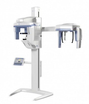
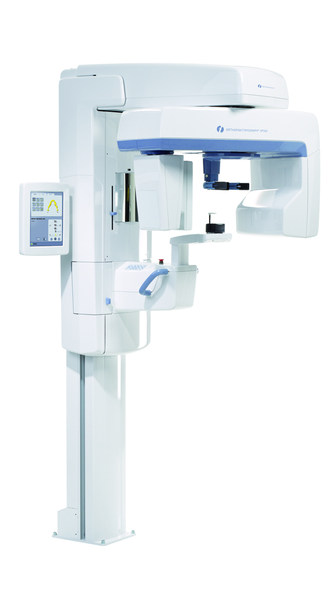
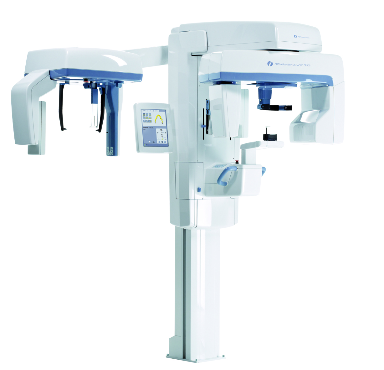
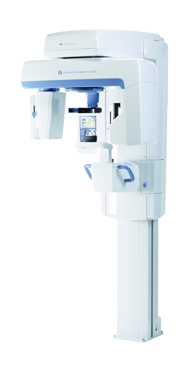
4Image quality is a result of many elements, such as the carefully planned features, chosen technology and sufficient technical characteristics of the system, along with proper patient positioning. The ORTHOPANTOMOGRAPH® OP300 combines all these for your benefit and provides you with a perfect image – every single time. ORTHOPANTOMOGRAPH® masters the details.
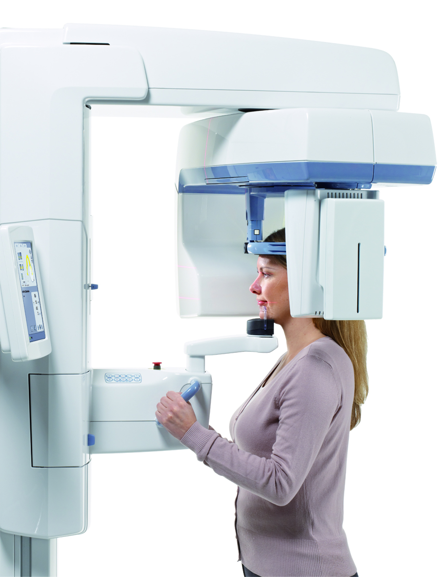
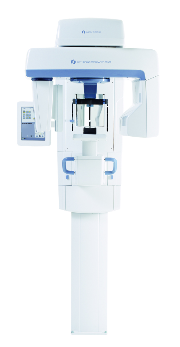
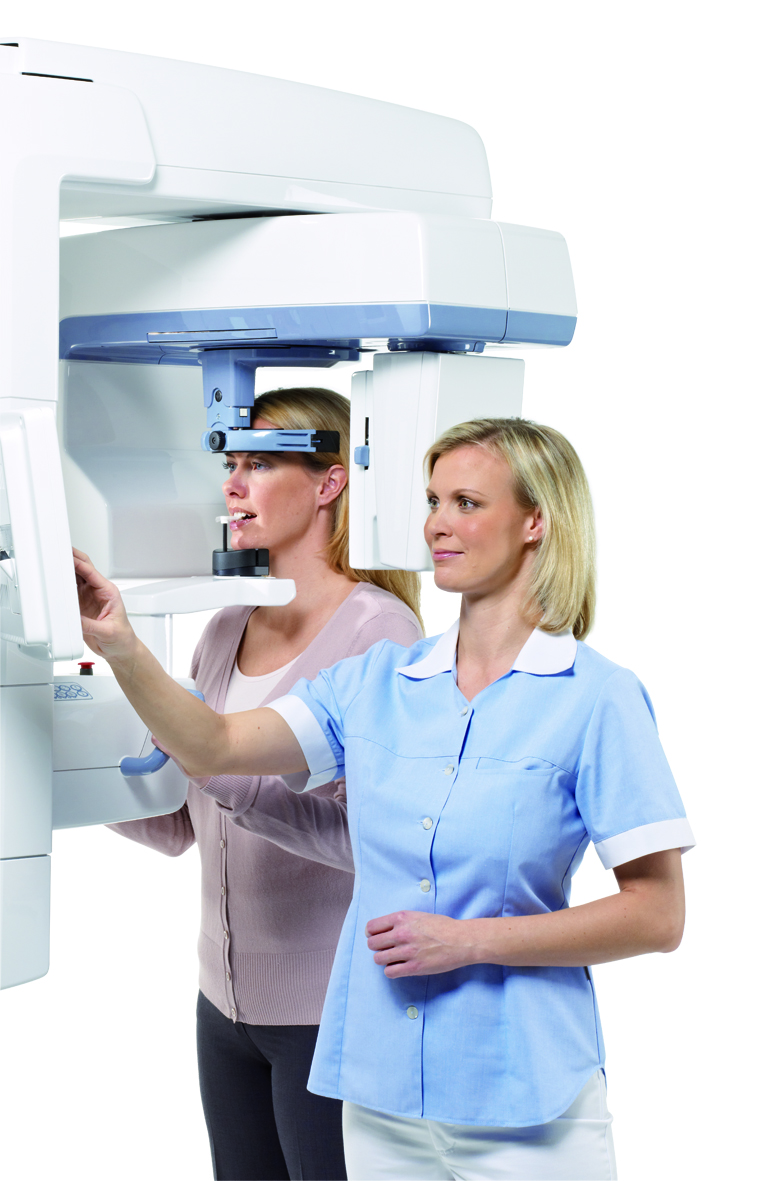
Stable and open patient positioning
A rigid 5-point positioning system, including forehead support, chin rest and bite fork, eliminates patient movement. The open design allows easy viewing and the positioning of the patient from either the left or right side.
Latest sensor technology
The OP300 utilizes the latest in CMOS sensor technology. CMOS sensors provide a larger dynamic range combined with 14-bit image data and increased signal-to-noise ratio. The result is intensely sharp image with reduction of unwanted under- and overexposures.
Complete usability
Large 10” touchscreen with easy-to-use interface enables professional usage from the very beginning. Clear and user friendly structure of controls allows fast and effortless workflow for all imaging modalities.
Multilayer pan
The OP300 multilayer panoramic option provides five panoramic images with only one scan. This enables forgiving patient positioning and reduces possible retake exposures. Multilayer images are achieved in the same scanning time and dose as the traditional panoramic scan.
Two available fields-of-view with 3D option
- 6 x 4 cm – a small FOV optimized for local diagnostics like single implant planning, 3rd molar extractions and endodontic procedures, keep the patient dose at a substantially reduced level.
- 6 x 8 cm – FOV covering complete dental arch for multiple implant placement and operations using surgical guides.
Two available resolutions
Both FOV sizes are high enough to easily cover the jaw bone and occlusion level. For both FOV sizes it is possible to choose between two resolutions:
- Standard scan takes only 10 seconds with exposure time only 2.3 seconds with optimized patient dose
- High resolution scan offers extremely sharp images for more detailed diagnosis
Metal artefact reduction (MAR) tool for OP300
Metal artifact reduction (MAR) tool can be used in 3D cases to reduce the effect of metal and other dense radio-opaque materials in the 3D image:
- Endo cases for better definition of root canals
- Post-op scans of implant cases
- All 3D scans of patients who have multiple fillings, bridges, braces or other metallic objects in their mouth
SMARTVIEW
A two dimensional scout image is taken before the 3D examination to adjust the target position visually from GUI screen. This guarantees precise positioning and eliminates risk of retake exposures.
- Select freely FOV position from GUI.
- Fine tune FOV position from GUI.
- SMARTVIEW ™ takes two dimensional scout of the selected area.
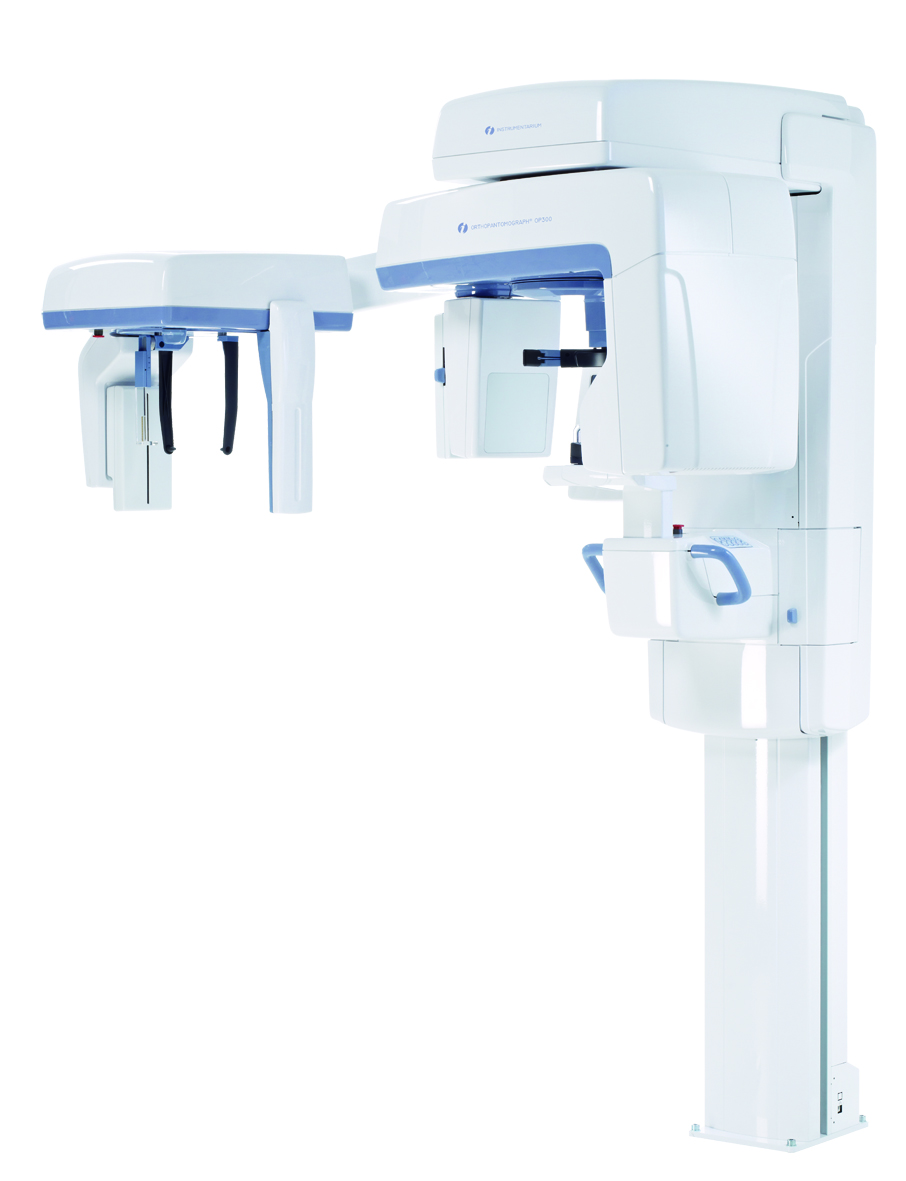
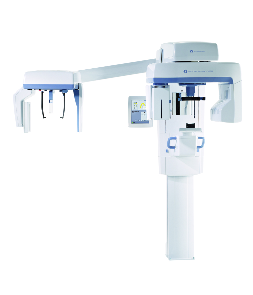
Unsurpassed cephalometric results
A scanning cephalometric option combined with a powerful X-ray generator and tube head offers unsurpassed visibility of tracing key reference points for orthodontic treatment planning. In addition, dose optimization is carried out by an adjustable scanning area and AFC (Automatic Facial Contour).
Fully adjustable scanning
Full range of projections: lateral ceph, AP/PA, obliques.
- Fully adjustable scanning area ensures that by exposing only the required region, patient safety is greatly increased.
- Automatic facial contour (AFC) decreases exposure factors in the facial soft tissue region to provide improved visibility of soft tissue tracing points in addition to a reduction in patient dose.
For more information please see the manufacturers website HERE