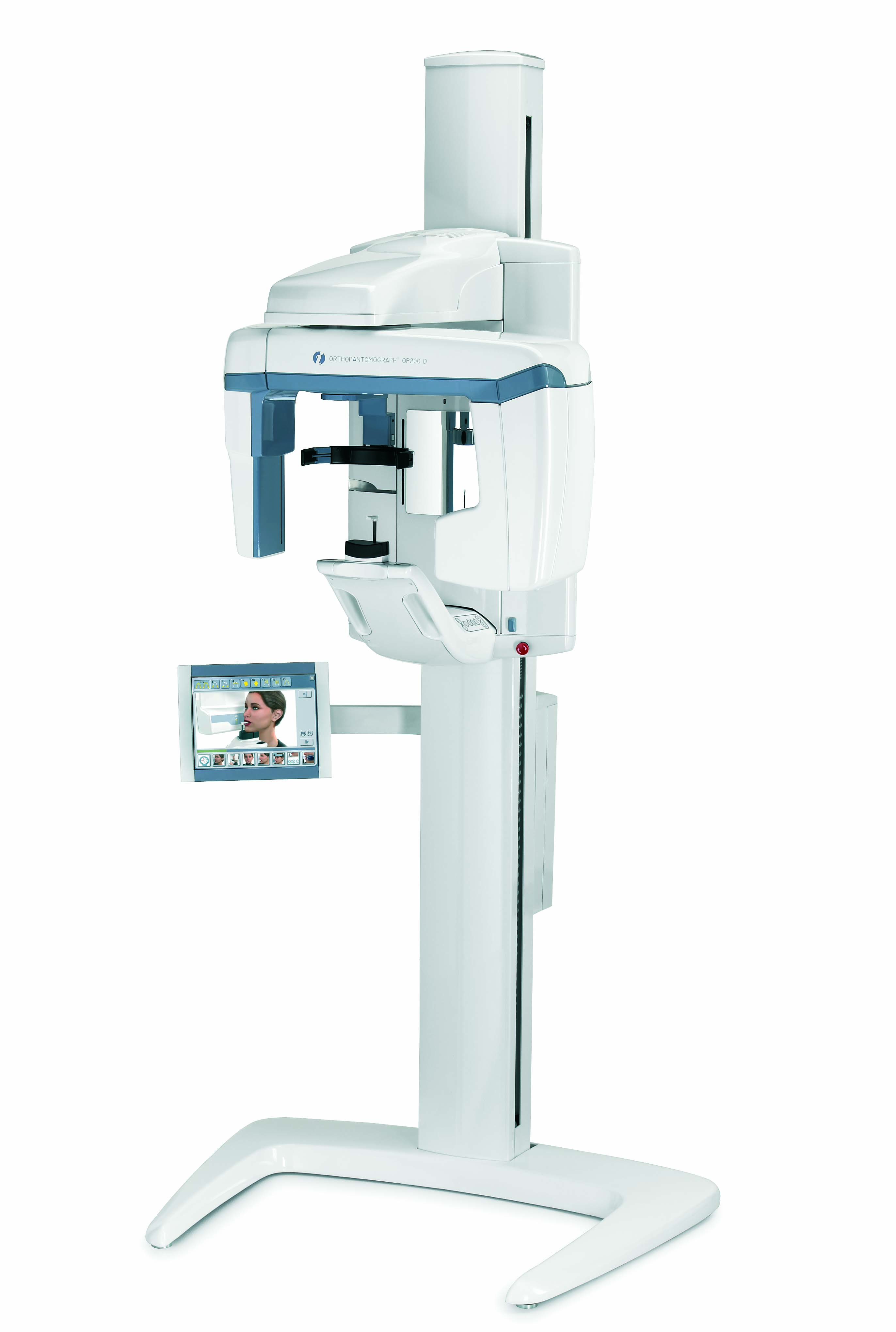
Correct imaging values – automatically
The OP200 D has a patented method for dose-controlled Automatic Exposure Control. The system measures patient bone thickness from the ramus and defines individual exposure values for patients with different sizes. This also enables individual Automatic Spine Compensation values to reduce spinal shadow in the image for each patient.
V-shaped beam – clinically proven imaging geometry
The V-shaped X-ray beam adapts to the human anatomy, providing even greater detail and a wider mandibular image layer. The V-shaped X-ray beam also allows for more penetrating power for the thicker maxilla area.
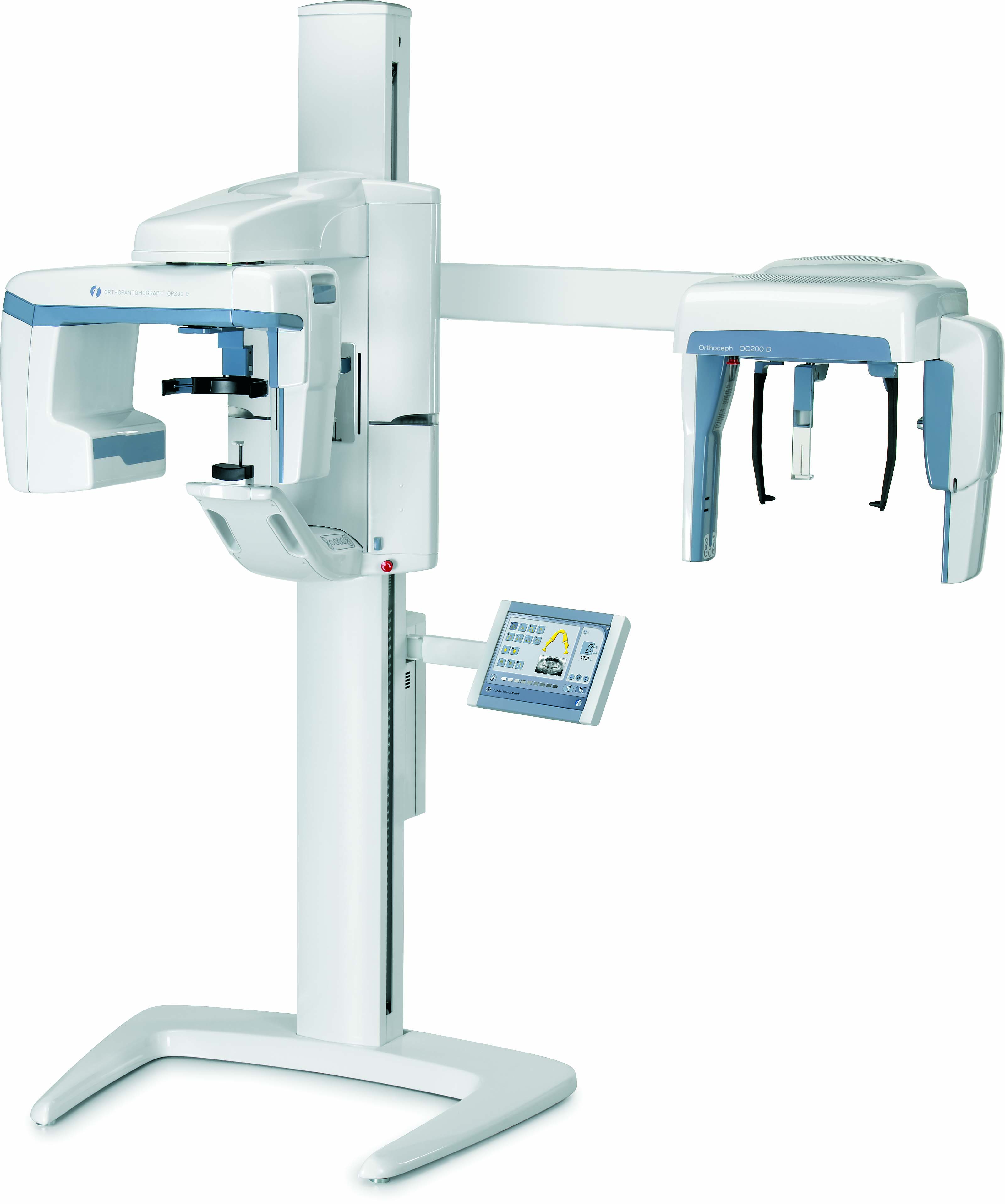
Fully adjustable scanning
The OC200 D incorporates an advanced user-adjustable lateral scan method to expose only the desired portion of the skull. This method reduces the scanning time to a minimum of 5 seconds and reduces patient dose considerably.
The OC200 D uses a patented Automatic Facial Contour (AFC) method for soft tissue enhancement in lateral views. The unit automatically adjusts the exposure values during scanning for better soft tissue definition.
48% to 62% dose reduction
Clinically correct image geometry
In order to produce equal and accurate horizontal and vertical magnification, the OC200 D uses a patented method of synchronized tube head horizontal sweep and sensor movements while keeping the focal spot in the same position.
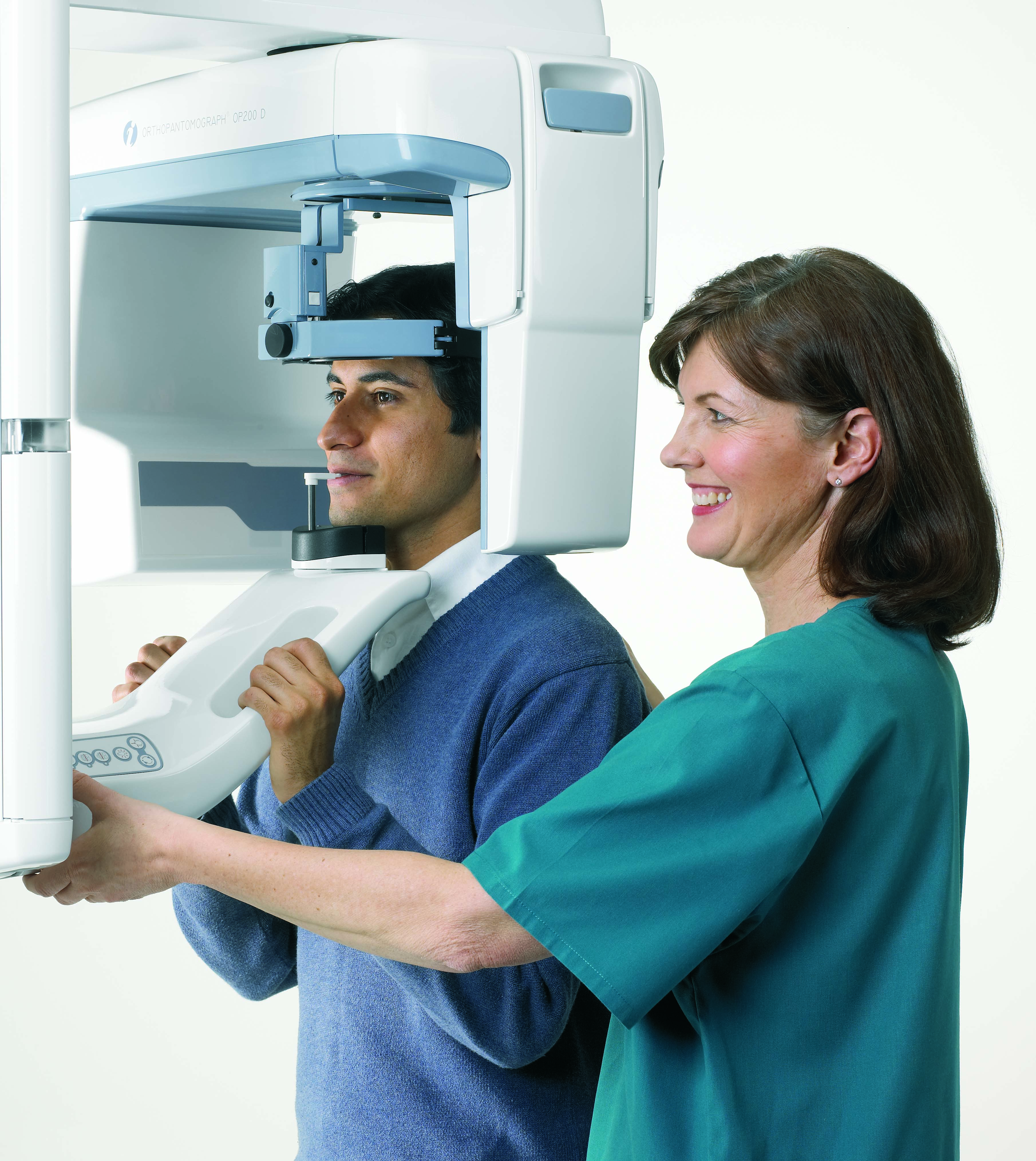
Stable patient positioning
The Frankfurt horizontal plane laser light, nasion support and rigid ear rods with locking system make patient positioning easy and convenient.
Perfect fit for your clinic
OC200 D can be set up in your clinic for right- or left-handed cephalometric imaging and is “field changeable” smartpad can be installed on either side of the unit or on the wall.
Full range of projections
The patient positioning system provides a variety of imaging projections for cephalometric radiography. It is a comprehensive diagnostic device that includes lateral, facial, posterioranterior and oblique projections, as well as the possibility of hand and wrist imaging.
The optimal solution
- Very easy and forgiving patient positioning
- No measuring of patient or marking of impressions required
- Upgradeable to every OP200 unit
The OP200 with VT is the most advanced and comprehensive cross-sectional imaging system on the market. It provides accurate and valuable information especially for implant planning. With the VT there is no need for unit modifications or purchasing expensive sensors.
Easy navigation of slices
The VT system has a slice navigator that shows the exact position of the cross-sectional slice in real time.
Excellent image quality
Our unique reconstruction method produces high-quality images using a patented method for making crosssectional slices with narrow X-ray beam and standard panoramic sensor. This has been proven to give better image quality than other known reconstruction methods.
Implant planning tools
The implant planning tool helps you to easily determine the correct implant for treatment. The tool contains implant models from leading manufactures. The software also provides the necessary measuring tools.